![]()
ROTH PHILOSOPHY
R05
Imaging CT & MRI
-

01R05
Assessing the Condition of the Temporomandibular Joints (TMJs)
The advent of CT and MRI has had a decisive impact on our Roth Study Club. In 2004, cone-beam computed tomography (CBCT)—a modality that exposes patients to relatively low radiation while providing high-resolution images—became available.
Furthermore, MRI has become widely accessible in Japan, allowing imaging to be performed at a lower cost compared with many other countries. This enables us to determine the presence of inflammation in the TMJs and to identify soft tissue abnormalities, particularly articular disc displacement.
TMJ problems can also occur in children prior to orthodontic treatment. In particular, assessing the status of the articular disc displacement is essential for confirming the stability of the TMJs.
-
TMJ CBCT Image
 Initial
 Post-splint
PDW MRI images
 Initial
 Post-splint
T2W MRI images
 Initial
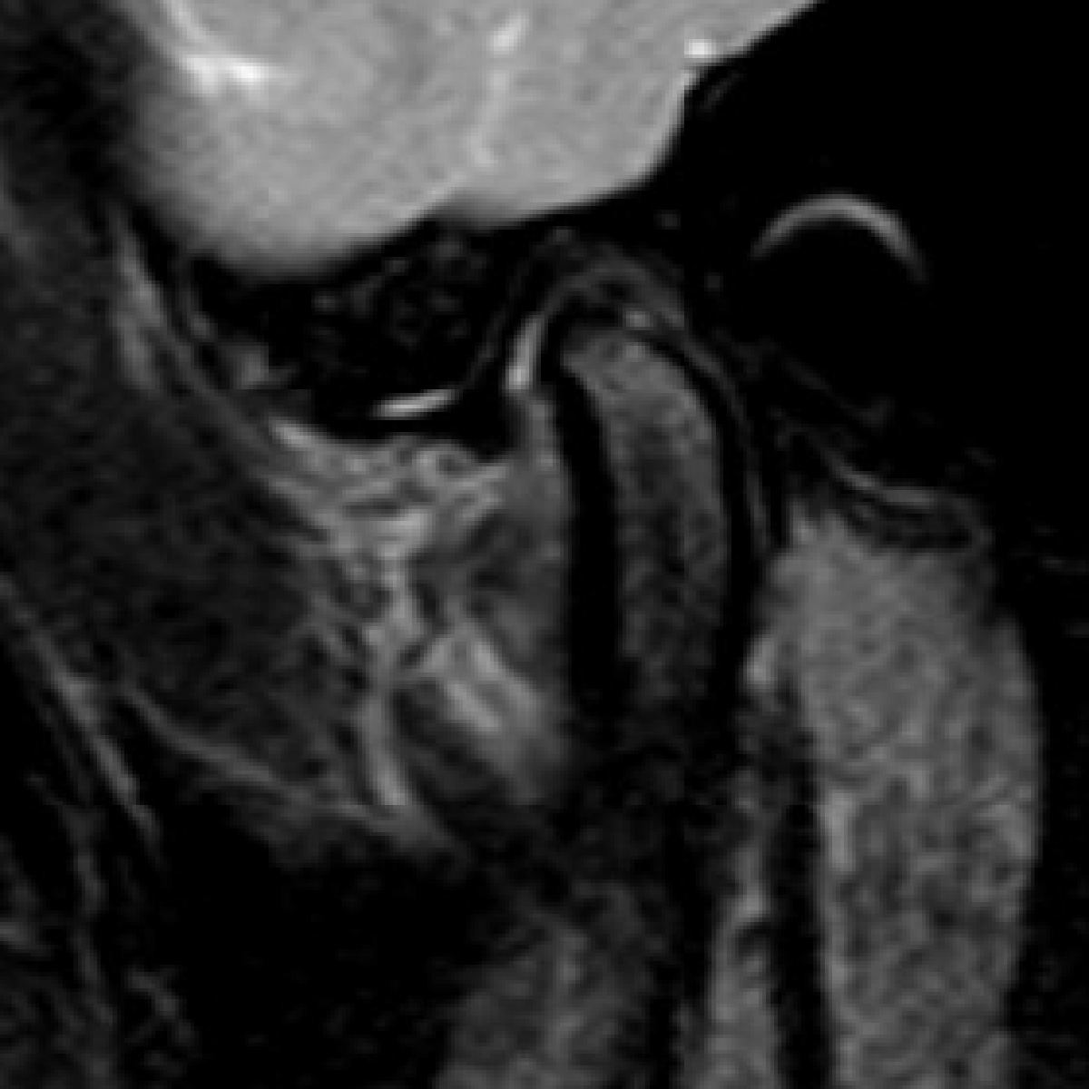 Post-splint
Intraoral photos
 Initial
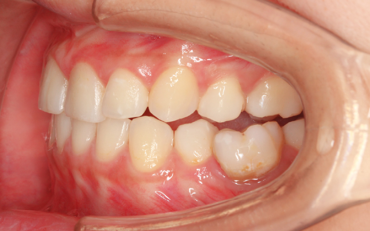 Post-splint
Case 01R05
A Case of Splint Therapy
TMJ CBCT Images
The position of the mandibular condyles has changed and is now closer to a normal position.
PDW MRI images
An improvement in the position of the articular disc can be confirmed.
T2W MRI images
The high-signal (white) area observed at the initial examination indicates inflammation. This is a common finding when disc displacement occurs. We often observe a reduction in this inflammatory signal following splint therapy.
Intraoral Photos
The intraoral occlusion has also changed, clearly demonstrating the close relationship between the condition of the TMJs and intraoral occlusion.
Although disc displacement that has progressed to this extent does not always improve, the favorable changes observed in this case were achieved because the patient wore the splint consistently for 24 hours a day.
-
TMJ CBCT Images
- Initial
- Post-splint
The position of the mandibular condyles has changed and is now closer to a normal position.
-
PDW MRI images
- Initial
- Post-splint
An improvement in the position of the articular disc can be confirmed.
-
T2W MRI images
- Initial
- Post-splint
The high-signal (white) area observed at the initial examination indicates inflammation. This is a common finding when disc displacement occurs. We often observe a reduction in this inflammatory signal following splint therapy.
-
Intraoral Photos
- Initial
- Post-splint
The intraoral occlusion has also changed, clearly demonstrating the close relationship between the condition of the TMJs and intraoral occlusion.
-
Although disc displacement that has progressed to this extent does not always improve, the favorable changes observed in this case were achieved because the patient wore the splint consistently for 24 hours a day.
Featureof the Roth Philosophy
-
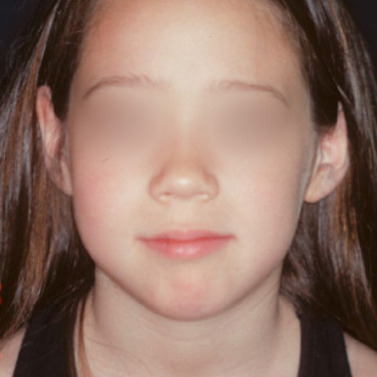
Before
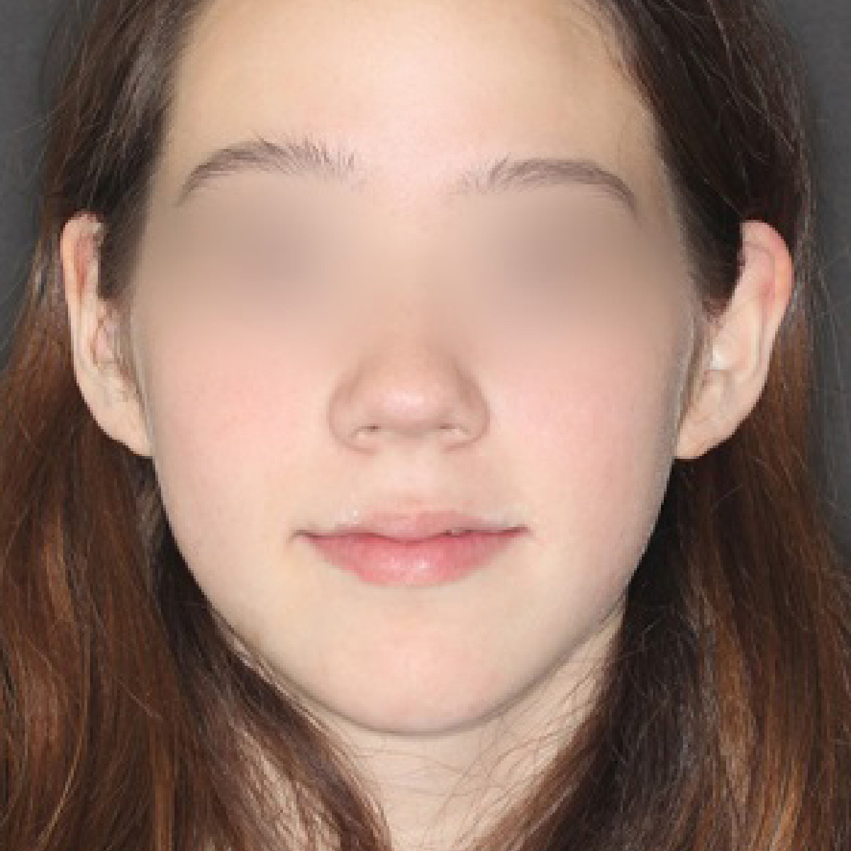
After


R01
Face Design
Strong Focus on Facial Impact
When considering the principal region of the face—from the eyebrows to the chin—the tooth crowns and roots account for nearly 40% of the area. Therefore, moving the teeth has a significant impact on overall facial appearance.
-


R02
Attractive Tooth Alignment
Attractive, Beautiful Tooth Alignment
Simply straightening the teeth does not automatically result in a truly beautiful tooth alignment. A well-formed dental arch, a stable mandibular position, and healthy, stable temporomandibular joints (TMJs) contribute to a beautiful smile and promote the health of the teeth and periodontium.
-


R03
Beautiful Smile
Lips and Tooth Alignment in the Smile
The relationship between the lips and tooth alignment is surprisingly important. It can make a significant difference in the appearance of the smile.
-

R04
Stable Jaw Position
The importance of mandibular position
While the alignment of the teeth is important, it is essential to first assess the condition of the TMJs. This approach is similar to conducting a soil survey before building a house.
-
R05
Imaging
CT and MRI
In addition to cone-beam CT, which offers high accuracy with low radiation exposure, MRI is widely available in Japan and can be performed at a lower cost than in other countries. This allows us to thoroughly assess the condition of the TMJs.
-
Before
After


R06
Optimum Timing
The Right Timing for Treatment
It is important to identify the stages of a child’s growth—particularly the growth of the mandible (lower jaw)—and to tailor the treatment plan and procedures accordingly.
-
Before
After
R07
Early Treatment
Early Treatment for Children
Early treatment is recommended for cases in which treatment would become more difficult later, or in which leaving the condition untreated would place a significant burden on chewing function and the TMJs.

